HAND X RAY PA HAND RadTechOnDuty
This diagnostic tool can help your doctor locate and understand injuries or degenerative diseases that affect one or both of your hands. Your doctor can also use hand X-rays to monitor the growth.
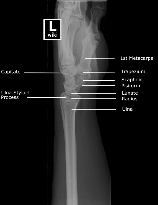
Wrist Radiographic Anatomy wikiRadiography
Diagnostic Labels for Musculoskeletal Pain. Appropriate diagnosis of musculoskeletal pain involves a multifactorial approach that includes the history of the disease, a thorough physical.

[Figure, Wrist xray with labeled osseous anatomy] StatPearls NCBI Bookshelf
Key points. Finger injuries visible on X-ray include bone fractures, dislocations and avulsions. The hand comprises the metacarpal and phalangeal bones. Fractures and dislocations are usually straightforward to identify, so long as the potentially injured bone is fully visible in 2 planes. Finger joints commonly dislocate and are susceptible to.

Read on to find out more about my review areas on a hand XRay... 👨🏽💻Hand xrays are
The wrist is one of the most commonly requested X-Rays in the children's emergency department. Wrist views are requested when injury to the distal radius/ulna or carpal bones are suspected.

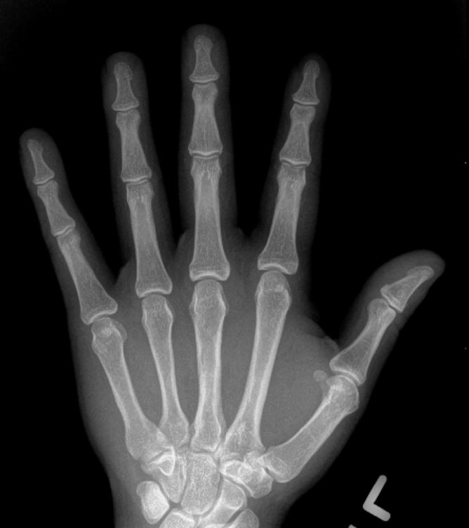
Normal Hand X Ray Colorvir Xray photo of normal right hand Stock Image Find the
Description. Hand X-Ray Anatomy and Interpretation Checklist 1. Soft tissues - Look carefully at the soft tissue over all the bones for any swelling or foreign body. The swelling should prompt a careful search of the underlying bone or joint.⠀ 2. Bones - All the bones of the hand should be examined carefully and systematically.

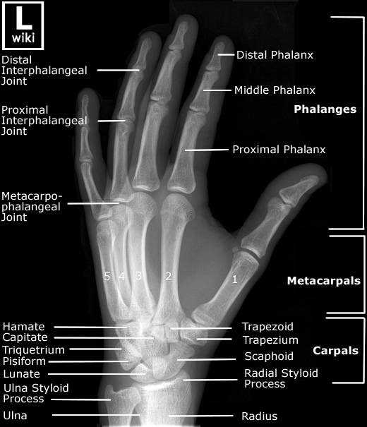
Hand Radiographic Anatomy wikiRadiography Radiology student, Radiology schools, Medical anatomy
Citation, DOI, disclosures and article data. The hand series consists of posteroanterior, oblique, and lateral projections. Although additional radiographs can be taken for specific indications. The series primarily examines the radiocarpal and distal radioulnar joints, the carpals, metacarpals, and phalanges.
What does hand arthritis look like on xrays? Raleigh Hand Center Raleigh Hand Center
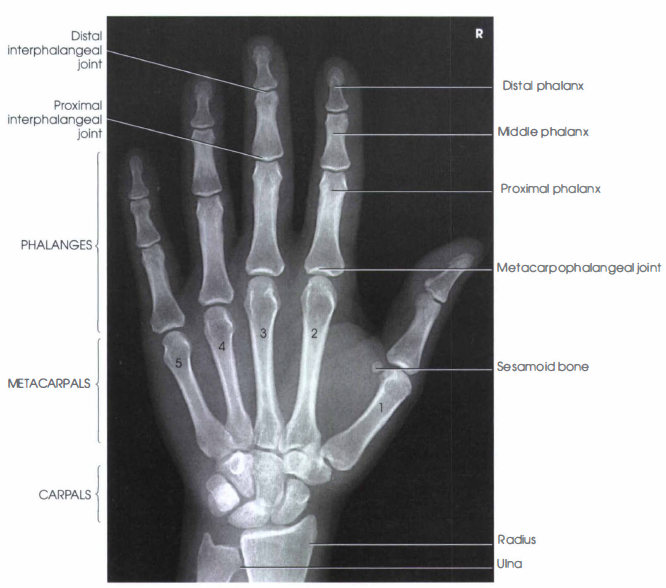
Shaft of third metacarpal. Neck of fifth metacarpal. Head of forth metacarpal. Metacarpophalangeal joint. Proximal phalanx. Middle phalanx. Distal phalanx. Sesamoid bones (flexor pollicis brevis, adductor pollicis). Terminal tuft.

Hand Radiographic Anatomy wikiRadiography Diagnostic imaging, Medical knowledge, Medical anatomy
extends from the radiocarpal joint to the tips of fingers. similar series. wrist series. distal radius and ulna, carpals and proximal metacarpals. scaphoid series. wrist series plus two additional scaphoid views. thumb series. just for looking at the thumb. both hands.

Xray Hand
A hand X-ray (radiograph) is a test that creates a picture of the inside of your hand. The picture shows the inner structure ( anatomy) of your hand in black and white. Calcium in your bones absorbs more radiation, so your bones appear white on the X-ray. Soft tissues, such as muscle, fat and organs, absorb less radiation, so they appear.
What a normal hand xray looks like (left) and what I managed to do to my hand(right). Local
Hand X-ray Guideline. Routine: 3 views • PA • PA OBLIQUE • LATERAL - Separate fingers to prevent overlapping (Fan lateral) Foreign Body: 2 views • PA
Hand Radiographic Anatomy wikiRadiography
A hand x-ray is taken in a hospital radiology department or your health care provider's office by an x-ray technician. You will be asked to place your hand on the x-ray table, and keep it very still as the picture is being taken. You may need to change the position of your hand, so more images can be taken.
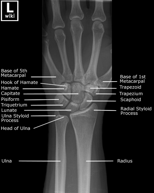
Wrist Radiographic Anatomy wikiRadiography
Access my FREE Online Membership today → https://www.thenotedanatomist.com___Unlock my Premium Tutoring Memberships → https://www.thenotedanatomist.com/premi.

Frontal view x ray bones hand Royalty Free Vector Image
A physician may perform a hand x-ray, MRI or ultrasound to rule out, assess, evaluate and diagnose the problem. A hand x-ray is often used to determine type of injury, extent of injury, and helps to determine treatment of the injury. Hand x-rays can detect broken bones and arthritis of the hand.

Pin on Xrays
Why the Test is Performed. Hand x-ray is used to detect fractures, tumors, foreign objects, or degenerative conditions of the hand. Hand x-rays may also be done to find out a child's "bone age." This can help determine if a health problem is preventing the child from growing properly or how much growth is left.

Normal Hand X Ray Colorvir Xray photo of normal right hand Stock Image Find the
TSA Cares is a resource that provides travelers with disabilities, medical conditions, and those that need additional assistance with information to better prepare them for the security screening process. In addition to the assistance, TSA has modified procedures to ensure that your screening experience is smooth and seamless.
Causes and Management of Wrist Joint Pain Complete Orthopedics
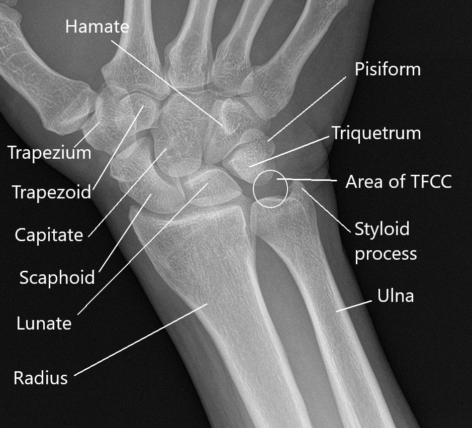
Wrist x-ray with labeled osseous anatomy. The .gov means it's official. Federal government websites often end in .gov or .mil.